Posts on chest x-rays do not show likely impact of COVID-19
- This article is more than six years old.
- Published on May 18, 2020 at 23:42
- 3 min read
- By Louis BAUDOIN-LAARMAN, AFP Canada
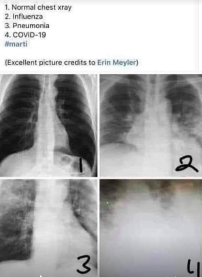
“Maybe staying home is clearer to understand now,” the caption of a Facebook post shared thousands of times since April 2020 says. Below it, four numbered x-rays show a human chest cavity in different conditions.
The first photo shows a clear chest, while a white fog covers parts of the thorax in the second and third images, which are said to show influenza and pneumonia-afflicted chests.
In the fourth image, which claims to show the x-ray of a patient with COVID-19 -- the disease caused by the novel coronavirus -- the chest is almost entirely hidden behind a dense white fog.
Screenshot of a Facebook post taken on May 15, 2020
Although most versions of the posts on Facebook and Twitter credit Erin Meyler, Meyler shared an April 22, 2020 post by Glenn Levine, a nurse in New Jersey, who appears to be the first to have shared the images.
Levine did not answer requests for comment by the time of publication.
Experts told AFP the photos can represent the conditions mentioned in the posts, but that the majority of COVID-19 patients do not experience lung damage as drastic as that shown in the fourth photo.
“Most COVID-19 cases do not look like (photo) #4. However, it is possible #4 is from a patient with severe COVID-19,” John Eng, a professor of radiology at John Hopkins University’s School of Medicine, told AFP by email.
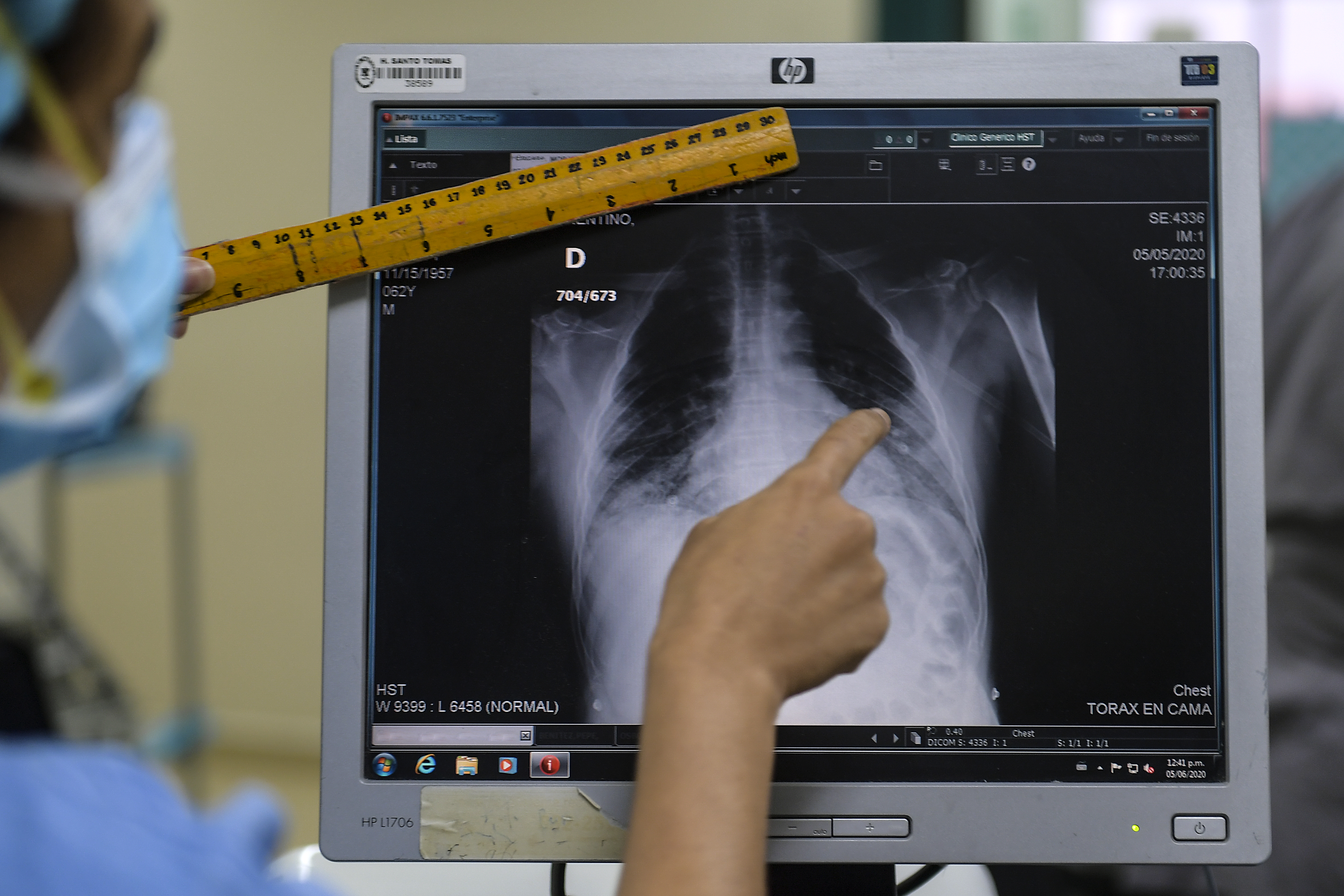
Gary Horn, a radiologist at the Baylor College of Medicine, said by phone: “It seems misleading. Any one of these four chest x-rays could be COVID.”
Horn explained that findings on chest x-rays, like the opacity in the photos, can represent a variety of things, such as accumulations of abnormal fluid, pus, blood, or cells.
“Anything that blocks the x-rays from getting through the body is going to be white,” he said.
Arash Bedayat, a radiologist at The University of California, Los Angeles, said by phone that “even in the most severe cases of COVID, in the first 48 hours, the chest x-ray can be completely negative.”
Bedayat, who works daily with COVID-19 cases, pointed out that some patients with the novel coronavirus may not show any opacity on a chest x-ray. Other asymptomatic patients who came in for different treatments had x-ray findings that were later linked to the virus.
In confirmed cases of the novel coronavirus, 58.3 percent of chest x-rays were normal, according to an April 2020 study published in the Journal of Urgent Care Medicine that looked at 636 patients.
Both Bedayat and Horn said that in chest x-rays from COVID-19 patients that do show signs of the disease, the opacity is often found in the lower zone of the chest, and away from the heart.
This is consistent with the findings of a March 2020 study from Hong Kong’s Queen Mary Hospital that looked at 255 chest x-rays of COVID-19 patients.
The study includes examples of chest x-rays from novel coronavirus patients, none of which look like the fourth photo in the online posts.
“It does matter because it’s wrong,” Bedayat said of the post, calling it “a bad image for a good cause.”
“The good aspect I see in that image is that it emphasizes that this disease is serious and we should make sure we do everything not to get it, because it can get very bad,” Bedayat said.
AFP Fact Check has debunked more than 450 examples of false or misleading information about the novel coronavirus crisis. A complete list of our fact checks on the topic in English can be found here.
Copyright © AFP 2017-2026. Any commercial use of this content requires a subscription. Click here to find out more.
Is there content that you would like AFP to fact-check? Get in touch.
Contact us